
Medical care
Ultrasound
Abdominal organs, thyroid, breast, blood vessel and joint examination
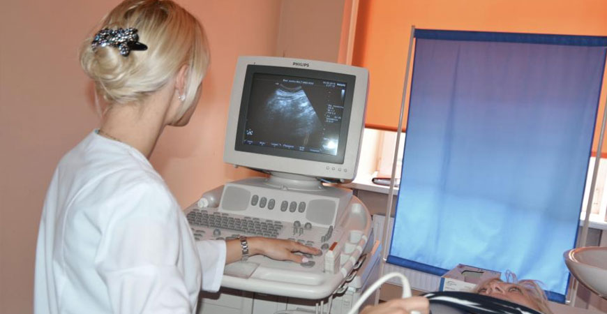
Ultrasonography is a non-invasive, painless, safe and fast examination method, which enables the diagnosing doctor to evaluate the condition of your internal organs and tissues. During the examination the patient is not irradiated, as the method is based on ultrasound waves reflecting off the tissues and the signal being visualised on the screen. The method is also used to evaluate pathological conditions and formations, as well as the treatment process over time.
Abdominal organs
During the examination the liver, gall bladder and bile ducts, pancreas, kidneys and urinary system are assessed, along with the main blood vessels of the abdominal cavity, the lymph nodes and the intestines.
Recommendations before the examination:
- For 1–2 days before the examination follow a diet and avoid foods that cause flatulence (bread, milk, cabbage, fresh vegetables and fruit, sweets).
- Do not eat for at least 4 hours before the examination.
- If possible, do not empty the bladder before the examination.
Small pelvic organs
During the examination the small pelvic organs (bladder, prostate, and uterus with ovaries), the lymph nodes and the blood vessels of the small pelvis are assessed.
Ultrasound of the thyroid gland
This method allows the size (volume) of the gland, its structure and blood supply to be estimated, as well as pathological changes to be identified — the presence of nodes or cysts, their size and structure. The examination is necessary to establish an exact diagnosis, to choose the method of treatment and to monitor treatment over time.
Ultrasound of the mammary glands
An examination method during which benign or malignant formations of the mammary glands are identified, as well as other pathological processes (inflammation, abscess). The method is harmless and is used both independently and in combination with mammography. It is recommended as a preventive check-up for the early detection of breast cancer.
Ultrasound of joints and muscles
The method makes it possible to determine pathological processes in the joints associated with age-related changes or traumatic injuries: damage to the structure of the joints, the articular capsule, the ligaments and the cartilage. It also helps in diagnosing inflammatory processes and determines the presence of fluid in the joint. Unlike X-rays, the method is harmless and also allows the effectiveness of treatment to be monitored.

Contacts
Book an appointment
Call or write — our administrators will help you choose the specialist that matches your complaints.